Microbiology:
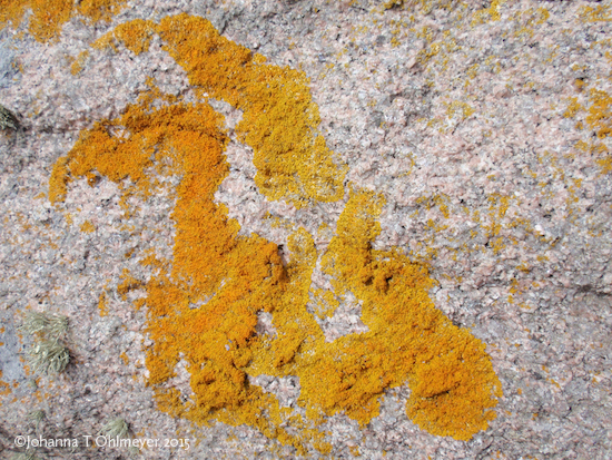
Bacteria form social communities that organize biofilm production: Most bacterial species have the ability to form colonies capable of creating biofilms. Biofilm formation is of medical concern because it confers the ability to adhere to surfaces and offers protection to pathogenic bacteria form chemicals such as antibiotics. These ecosystems can consist of a monolayer or multilayer of bacterial cells embedded in extracellular matrix composed of polysaccharides, proteins, and DNA. Bacteria within the biofilms communicate via quorum sensing; this is important to control the rate of reproduction and amount of secreted extracellular material. Once a critical mass is reached, dispersal to a new site occurs. Biofilm growth depends on the availability of carbon and nitrogen resources for all bacteria in the biofilm, cells in the inner part of the biofilm as well as the outer part or peripheral bacteria. Since nutrients from the environment will reach the bacteria at the periphery first, the inner bacteria need to compete for nutrients. On the other hand, the outer bacteria need to cooperate with their inner peers because if there is a mechanical or chemical (antibiotics) insult, the first to die are the peripheral bacteria. Regrowth of the biofilm will depend on reproduction of the inner bacteria. A paper by Liu et al. (2015) reports on efforts to elucidate the mechanism of this cooperation-competition interaction that regulates Bacillus subtilis biofilm growth.
The main observation is that biofilm size increases smoothly until a threshold is reached, after which biofilm growth oscillates with periods of increase rate of cell division followed by reduction of growth. Interestingly, the oscillatory growth only happens in the peripheral cells (Liu, et al., 2015). The authors hypothesized that the growth oscillations may be due to nutrient limitations specifically sources of carbon and nitrogen. Supplying carbon sources does not affect growth oscillations. Glutamic acid was supplied externally as a source of nitrogen, however the oscillations stopped only when glutamine or ammonia was supplied as an external source of nitrogen. Glutamine synthetase (GS) uses glutamic acid and ammonia as substrates to make glutamine. Glutamic acid from the media that bath the cells is readily available to the peripheral bacteria in the biofilm; while the inner bacteria have to compete for this nutrient. Ammonia is produced within the biofilm by the inner cells and diffuse out toward the periphery. The hypothesis is that as the peripheral cells reproduce, glutamic acid from the media is used up and the inner cells starve. This causes the inner cells to produce less ammonia. The peripheral cells reduce the rate of division because they do not have ammonia to make glutamine (the valley of the oscillation). Reducing the outer cells rate of division allows more glutamic acid to reach the inner cells, the renewal of ammonia production, and the increase of peripheral cell reproduction (the peak of the oscillation).
In order to test this hypothesis and make testable predictions and questions, the authors made a mathematical model, which mirrored the experimental observations. One obvious question is why the outer cells do not produce their own ammonia? Ammonia is produced from glutamate by the Glutamate Dehydrogenase enzyme (GDH), and the peripheral cells have enough external glutamate. One hypothesis is that the peripheral cells have to cooperate to keep the inner cells alive. By not producing their own ammonia, these cells are dependent on the metabolism of the inner cells and thus establish a nutrient co-dependence that acts as a sensor. In order to test this hypothesis the cells were treated with hydrogen peroxide or chloramphenicol, the peripheral cells die and growth of the inner cells increased. Thus, the bacterial cells in the biofilm form a community that organizes their metabolic activity in time (growth oscillation) and space (inner and outer cells). The goal is to cooperate to keep all the cells alive by regulating the competitive used of nutrients and they do this by establishing negative feedback loop characterized by co-metabolic dependency.
How can I use this post for class instruction? One idea is to have your students read the post, identify and list the observations, from this information students should be able to draw the oscillations (using time in the x-axis and growth in the y-axis) and a diagram of the negative feedback loop showing the metabolic dependence of the inner and outer cells. Then the teachers could ask, “what if questions”, different ways to test predictions, what to do next, etc. The students should engage in discourse to voice and explain their answers.
If you want to write a lesson plan, have questions & suggestions? Ask the Owlet.
Bloom Taxonomy: Knowledge, comprehension, application, analysis, synthesis.
NGSS connections: HS-LS1 From molecules to Organisms. Structures and Processes HS-LS2: Ecosystems: Interactions, Energy, and Dynamics.
Crosscutting concepts: Cause and effect, systems and systems models, stability and change.
Practices: constructing hypothesis and designing solutions. Developing and using models. Engaging in discourse.
Stem cell Biology
Exclusive cell communication between stem cells and their niche cells is assured by novel structures called nanotubules. Stem cell’s self-renewal and viability depend on signals send by the surrounding cells or niche cells. It has been observed in different systems that stem cells are in close contact with the niche cells. Germline stem cells divide asymmetrically into another stem cell, located in apposition to the niche cells, and a daughter cell that will go on and differentiate. Thus after the first division these cells are different. The signals of self-renewing and maintenance that keep the stem cells identity, emanate from cells in the niche and must only reach the stem cell. These signaling proteins like the morphogen Dpp, are secreted into the extracellular space. The question is how the movement of these diffusible proteins is restricted so that they reach only the stem cell and not the adjacent differentiating daughter cell?
An elegant paper by Inaba and collaborators shows that the stem cells send out finger-like projections, called microtubule dependent (MT)-nanotubes, into the niche cells. The scientists use live imaging techniques to study how the MT-nanotubes assured specificity of self-renewal signals in the Drosophila melanogaster male germline stem cells. The analysis of MT-nanotubes was done in vivo because they are sensitive to fixation, this may explain why they have not been detected in the past. The MT-nanotubes are novel structures structurally and functionally different from flagella or cytonemes. The MT-nanotubes are only formed during the G1 phase of the cell cycle, after stem cell division. During cell division, mitosis, they are not detected. MT-nanotubes are finger-like projections containing microtubules surrounded by plasma membrane. Genetic analysis shows that mutating genes required for microtubule transport, reducing the amount of tubulin or depolymerizing it, negatively affect the formation of the projections. Conversely, stabilizing microtubules by adding Taxol or mutating a MT-depolarizing kinesin, increases the thickness and length of the MT-nanotubes. In addition, several proteins required for transporting materials up and down the microtubules have been observed along the MT-nanotubes by tagging them with GFP (green) or Cherry (red) fluorescent tags. Mutations on these genes affect the thickness and length of the MT-nanotubes.
A follow-up question is how exactly the MT-nanotubes restrict signaling between the stem and niche cells? Dpp is a well-characterized self-renewal signal and it is secreted from the niche cells to the extra cellular space. Since the MT-nanotubes protrude and form deep indentation in the niche cell, the point where signaling occur may be the tip of the MT-nanotubes. The authors clearly showed that the Dpp receptor Tkv (they use tagged Tkv-GFP) is observed along the microtubules and in the membrane at the tip of the MT-nanotubes. This suggests that Dpp binds its receptor Tkv at the tip of the projection and that the receptor localization to the tip of the MT-nanotube is regulated. A corollary of this result is that Dpp signaling may have a role in the formation and maintenance of the projections. Indeed, using a dpp temperature dependent mutant, Inaba et al. showed that decreasing the amount of Dpp signaling affect the formation of the MT-nanotubes. This paper shows how the interaction between the stem cell and the niche establish a close, specific connection in order to keep the self-renewal signal restricted. One follow-up question is how the binding of Dpp to Tkv at the tip of the MT-nanotubes generates a signal that is transduced to the stem cell so that it regulates the formation and maintenance of the MT-nanotubes?
Ecology
The symbiotic relationship among corals, algae, and the marine microbiome is central for the health of coral reefs. The calcium carbonate that forms the skeleton of coral reefs is built by the animal part, the coral, and its endosymbiotic partner, the photosynthetic algae of the genus Symbiodinium. The coral acquires enough nitrogen and other nutrients from heterotrophic feeding, when there is enough to eat. It also gets extra nutrients from the endosymbiotic algae in the form of photosynthates; fixed carbon products like aminoacids. However, these products are high in carbon and do not provide enough nitrogen. The algae are very effective in utilizing dissolved inorganic nitrogen (DIN) form the salt water in the form of ammonia and nitrate. The nitrogen is stored in the algae, used as needed, and pass to the host. The algae, in return, benefits from the relationship by receiving inorganic nutrients produced as coral metabolic waste.
The growth and density of the photosynthetic algae is highly dependent on the availability of nitrogen and these sources vary seasonally and daily. The question is how the coral regulates its symbiotic partner and affect nitrogen cycling so the coral reef has steady amounts of nitrogen. The coral controls algae growth in several ways; by releasing factors that induce the released of algae photosynthetic fixed carbon compounds, it also controls algae numbers by degradation or digestion, and it controls algae density by limiting nutrients. Limiting nitrogen availability to the algae is important because if surplus nitrogen is available the result is metabolic unbalance that leads to an increase on the Nitrogen to Phosphate (N:P) ratio, coral phosphate starvation, loss of their symbionts, and coral bleaching. However the long held idea that coral reefs are composed of coral and algae is incomplete because it is becoming clear that the coral reefs depends heavily on microbes, mainly bacteria, archaea, fungi, and virus. Together the coral, algae, and microbes form a complex ecosystem that is very effective in using and recycling nutrients. We need to know more about these complex interactions in order to understand how coral reefs respond to challenges like ocean acidification, increased ocean temperatures and disease.
One hypothesis is that coral reefs adapt to environmental conditions by regulating their microbiome, specially the abundance and type of microbes ( Radecker, Pogoreutz, Voolstra, Wiedenmann, & Wild, 2015). There are different type of microbes associated with diverse parts of the coral reef like, the mucus layer, the coral tissue, the coral skeleton, and so forth. We do not know the specific role of these microbes in their habitat; potentially they could assist with functions like nitrogen and carbon fixation, sulfur cycling, antimicrobial defense and others. However, it is clear that nitrogen cycling is important for the health of coral reefs and that the ubiquitous nitrogen-fixing microbes in the coral reef may be essential. Some aspects of the nitrogen cycle in coral reefs are being studied. Coral reefs are a high source of nitrogen fixation (N2 to ammonia), however the coral itself seems to fix very little nitrogen. There is high amount of nitrogen-fixing bacteria or diazotrophs associated with the coral reef and they are important for nitrogen assimilation. However, since nitrogen fixation utilizes 16 mols of ATP to fix 1 mol of N2, this mode of utilizing nitrogen may be only used when other sources of nitrogen are scarce. Another aspect of nitrogen cycle is nitrification. Nitrification, the conversion of ammonia to nitrite or nitrate is ubiquitous in the tissues of the coral reef, and there is abundant nitrifying bacteria and archaea in the coral reef skeleton and other tissues. However we do not know what is the function or role they play in the maintenance of the reef. One possibility is that these bacteria take away the ammonia form the algae by converting it to nitrate; algae prefers the use of ammonia to other forms of nitrogen. Thus by doing this the coral keeps low the amounts of nitrogen available to the algae and maintains metabolic balance. There are other aspects of nitrogen cycle like denitrification and the role of eukaryotic coral associates that needs to be investigated and the results incorporated to the nitrogen cycle in coral reefs.
Environmental conditions such as elevated temperatures and dissolved organic compounds can lead to nutritional imbalance with a net increase in the N: P ratio. This would destabilize the coral-algae symbiosis because the coral control of nutrients available to its symbiotic algae would be disrupted. The elevated availability of nitrogen would promote algae growth and further phosphate depletion. It has been shown that phosphate starvation causes the increase of sulfo- to phospholipid ratios, destabilization of thylakoid membranes, and coral bleaching. This area of coral research is important because the breakdown in coral-algae symbiosis can lead to the demise of coral reefs colonies that can expand to nearby colonies and eventually to whole coral ecosystems.
Medicine
A study reports the development of a promising new drug that targets all the life cycle stages of Plasmodium falciparum. Malaria is a vector-mediated disease that kills many people worldwide, most of the cases being children and pregnant women in poor areas of the world. One problem in controlling this disease is that current medicines have developed resistance against the parasite Plasmodium falciparum. We are in dire need to develop drugs with novel modes of action, that can be given in a single or few doses, and that have activity against the different life stages of the malaria agent, P. falciparum. The life cycle of P. falciparum can be divided in three stages; a liver stage, a blood cell stage, and the mosquito stage. Baragana and collaborators report the development of a new drug DDD107498 that is active against these three stages of the P. falciparum life cycle. It can clear the parasite from the blood, prevent the infection of hepatocytes, and interfere with transmission to the mosquito. The study started by screening a chemical library for activity against blood-stage P. falciparum strains that were sensitive or resistance to other anti-malarial drugs. Upon identifying a promising compound, medicinal chemists performed chemical optimization of the compound that led to good pharmacokinetic properties and an increase in compound potency by 100 fold compared to existing anti-malarial drugs. Another good characteristic of this compound is that it is potent enough to be given as a single dose.
Further characterization of the compound DDD107498 in a mice model of malaria showed that it can clear the parasite by 90% from the blood in a short time (48hrs). In addition DDD107498 has activity against other life-cycle stages. The first organ that P. falciparum colonizes, after the mosquito bite, is the liver. Experiments in mice show that if the drug is administered before exposure or after the initial infection has been established, the parasite is fully cleared and there is no sign of parasites after 30 days. This suggests that this drug can offer chemoprotection by acting on the first site of infection, the hepatocyte, and clearing the liver stage parasite. Another stage in the life cycle of P. falciparum at which the drug acts is in the blood cell when the parasite produces gametocytes. These gametocytes (stage I to V) keep circulating in the host’s blood even after the symptoms have disappeared. Gametocytes V is infectious to the mosquito, once ingested they go on to form gametes. One important activity of DDD107498 is that, unlike many current anti-malaria drugs, it inhibits gametogenesis at the parasite blood stage, thus this drug interferes with the transmission of the parasite from host to mosquito.
The next step in the investigation was to identify the molecular target. Given that the drug is active at many stages in the life of the parasite, it suggests that the target may be a protein used in basal metabolism. In order to identify the DDD107498 molecular target, the authors provoke resistance by exposing the parasite to high levels of the drug. Once resistance was developed, genomic DNA was extracted followed by whole-genome sequencing to find out the location of the mutations that confer resistance. There were 10 different strains with mutations on the gene that codes for Elongation Factor 2 (EF2). This protein is central during protein synthesis; EF2 is an elongation factor that mediates the translocation of the ribosome along the mRNA during protein synthesis. The importance of EF2 in protein synthesis of all cells explains why DDD107498 is active against several life cycle stages of the parasite. EF2 is a highly conserved protein but it has enough amino acid differences between P. falciparum, mice and human EF2 that it does not affect protein synthesis in human or mice cells.
DDD107498 represents a promising development in the fight against malaria, however it is a matter of time that this drug also will develop resistance (as demonstrated in this study). Thus we need to constantly produce medicines with different targets and use them in combination with other drugs. This combination therapy would help reduce the chances or slow the development of resistance. DDD107498 estimated cost is one dollar per treatment, so it is important that any drug used as partner in the combination should also be cheap in order to be at the reach of people who needed.
How can I use this post for class instruction? This paper is very interesting because it shows step-by-step how this drug was developed and it illustrates how different type of scientists, chemist and biologists, interact and work together to advance this project. We suggest first familiarizing the students with the P. falciparum life cycle by having each group draw it in poster size paper. Each group could obtain a “what if questions” from the teacher, debated among peers, and present the answer to the class along with different ways to test predictions, what to do next, etc.
If you want to write a lesson plan, have questions & suggestions? Ask the Owlet.
Bloom Taxonomy: Knowledge, comprehension, application, analysis, synthesis.
NGSS connections: MS-LS1-3, MSLS1-5, and HS-LS1-2: From molecules to Organisms. Structures and Processes. MS-LS2-2 and HS-LS2-6: Ecosystems: Interactions, Energy, and Dynamics.
Crosscutting concepts: Cause and effect, systems and systems models, stability and change.
Practices: constructing hypothesis and designing solutions. Developing and using models. Engaging in discourse.
Works Cited
Radecker, N., Pogoreutz, C., Voolstra, C. R., Wiedenmann, J., & Wild, C. (2015). Nitrogen cycling in corals: the key to understanding holobiont functioning? Trends in Microbiology , 23 (8), 490-497.
Baragana, B., Hallyburton, I., Lee, M. C., Norcross, N. R., Grimaldi, R., Otto, T. D., et al. (2015). Anovel multiple-stage antimalarial agent that inhibits protein synthesis. Nature , 522, 315-320.
Inaba, M., Buszczak, M., & Yamashita, Y. M. (2015). Nanotubes mediate niche–stem-cell signalling in the Drosophila testis. Nature , 523, 329-334.
Liu, J., Prindle, A., Humphries, J., Gabalda-Sagarra, M., Asally, M., Ly, S., et al. (2015). Metabolic co-dependence gives rise to collective oscillations within biofilms. Nature , 523, 550-565.